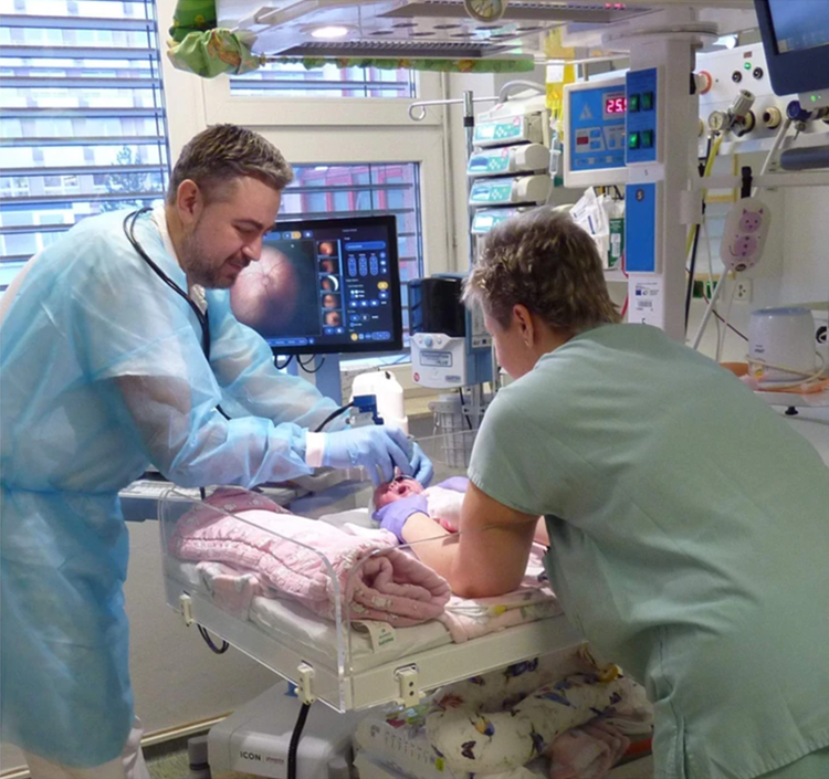
A groundbreaking advancement in the diagnosis and treatment of retinopathy in premature babies has been made by researchers in Ostrava. Using artificial intelligence, they are now able to detect serious retinal diseases more quickly and reliably, significantly reducing the risk of blindness in at-risk children.

PHOTO: Doctors at the University Hospital Ostrava performing retinal examinations on premature infants.
Retinopathy of prematurity (ROP), a disease affecting the retina of prematurely born infants, is the most common cause of severe visual impairment or blindness in children in developed countries. As a result, all premature babies undergo regular eye examinations, during which images of the retina are taken using specialized retinal cameras.
“We assess changes in the retina from these images, and if necessary, we administer a biological preparation directly into the eye or use lasers to treat the peripheral parts of the retina,” explains Juraj Timkovič, head physician at the Center for Children with Vision Impairments at the University Hospital Ostrava.
Currently, doctors rely on digital imaging systems to evaluate these retinal images, but these systems have limitations. To enable faster and more reliable diagnoses, healthcare professionals are collaborating with researchers from VŠB – Technical University of Ostrava to harness the power of artificial intelligence.
“Thanks to extensive screening conducted by doctors at the Ostrava University Hospital on premature infants, we had access to a dataset of 9,000 retinal images. In cases of retinopathy, pathological changes manifest as abnormal vascular curvature and changes in vessel thickness. These features served as key inputs for the artificial intelligence, which was trained to distinguish between healthy and damaged vessels,” explains Jan Kubíček from the Faculty of Electrical Engineering and Computer Science at VŠB-TUO.
Through advanced techniques, the researchers succeeded in developing algorithms and creating a highly accurate system for automatic segmentation of retinal blood vessels. This system aids in sorting images, simplifies the evaluation of retinal changes, and improves early disease detection.
“Artificial intelligence can help us not only diagnose specific stages of the disease but also assist with treatment. After administering medication, AI could determine whether changes have occurred and whether the blood vessels are starting to improve. It can thus help us predict the progression of the disease following treatment,” says Juraj Timkovič.
So how will the research findings be applied in practice? A web-based application is currently being developed, allowing users to upload retinal images for analysis by the system. “The application will primarily be accessible to medical professionals, enabling us to share information and experiences. Our main motivation is the practical use of our findings, which should improve diagnostics and increase the success rate of treatment in pediatric patients,” adds Jana Nowaková from the Faculty of Electrical Engineering and Computer Science.
The research is part of the LERCO project, a collaboration involving the University of Ostrava, VŠB – Technical University of Ostrava, and the University Hospital Ostrava. It is also part of the OP JAK cutting-edge research project Robotics and Advanced Industrial Manufacturing and the project Multi-Criteria Optimization with Constraints Using Potential Area Analysis (GF22-34873K).
Media coverage
